Brain-wide, MRI-guided electrophysiology
Recording prelimbic cortex and its inputs at once
Even simple behaviors depend on many brain regions working together, but most experiments record from one region at a time. Studying how prelimbic cortex (PL) interacts with its inputs means recording them together, and the part of each input region that projects to PL is often small, around 500 µm across, and deep. In mediodorsal thalamus, for example, only the anterior portion sends input to PL.
Standard stereotaxic targeting, from landmarks on the skull, misses by a median of 430 µm at 4 mm depth, and the error compounds across probes. At that accuracy, the odds that seven probes all land in seven 500 µm targets are roughly one in tens of thousands.
With Yoni Browning, Josh Siegle and Karel Svoboda, I adapted an approach from primate neuroscience to the far smaller mouse brain:
- Image each mouse with MRI before surgery (100 µm, 14 T). Register the scan to a population template and the Allen Common Coordinate Framework (CCF), so targets are planned in that animal’s own anatomy.
- Plan multi-probe Neuropixels insertions through a custom implant, avoiding collisions between probes, manipulators and hardware (Rutter).
- Record from many targets at once.
- Clear and light-sheet image the brain, register it to the CCF (iDISCO template), and trace each dye-labeled probe track (electrode tracer) to confirm where every recording site landed.
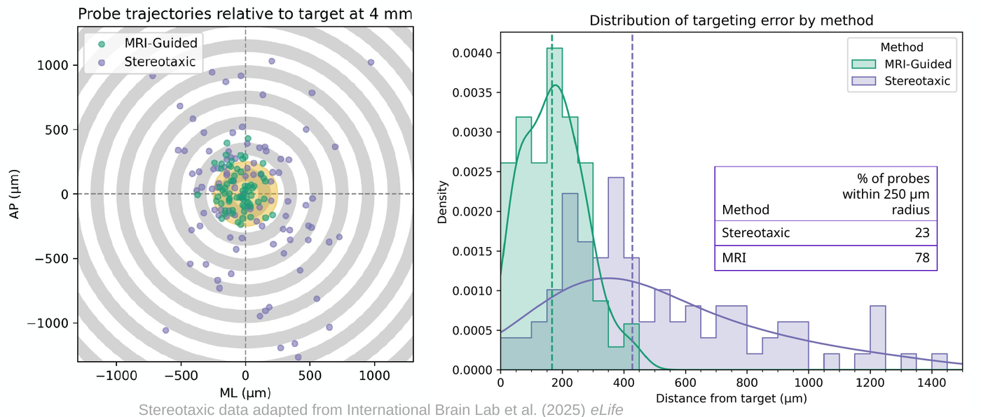
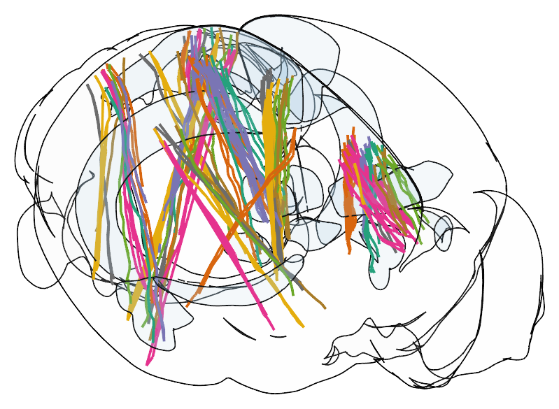
At this accuracy, a session has even odds of landing all seven probes in seven regions 590 µm across, where skull-based targeting would succeed about once in 3,900 tries. We have recorded 15 mice in 28 sessions, each targeting PL and six of its major inputs (mediodorsal and ventromedial thalamus, claustrum, basolateral amygdala, retrosplenial cortex and ventral CA1) while the mice perform a foraging task. Some sessions record up to fifteen 384-channel Neuropixels streams at once.
A manuscript describing the method is in preparation.